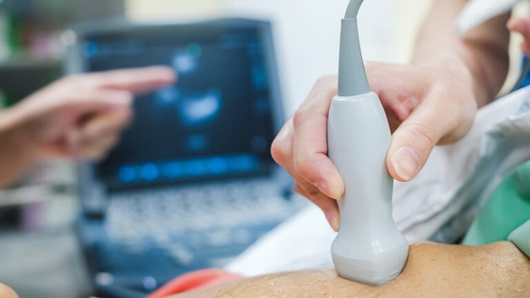
Artificial intelligence (AI) can assist in the creation of echocardiograms faster and more efficiently, improving image quality and reducing operator fatigue, the first prospective randomized control of AI-assisted echocardiography shows. It was shown in the test.
The Japanese study used Us2.ai software, developed from research platforms from 11 countries and supported by the Singapore Agency for Science, Technology and Research. This system and another newly developed AI system, PanEcho, developed at Yale University School of Medicine and the University of Texas at Austin in New Haven, Conn., can automatically analyze a wide range of structural, functional, and views of the heart. Research on these two systems was presented at the American Heart Association (AHA) Scientific Sessions 2024.
“This is what happens when you refer a computer scientist to a cardiologist,” says David O’Young, M.D., a cardiologist at Cedars-Sinai Medical Center in Los Angeles, California. “This allows us to develop some really exciting new technologies,” said Ouyang, who was not involved in the published research but is working with Echo, another AI platform developed at Stanford University in Palo Alto, California. -Leading previous research on Net Dynamic, fractions proven superior to human interpretation of echocardiograms for left ventricular ejection.
AI speed and accuracy
Echocardiography, the most common form of cardiac imaging, is “an ideal place to use AI,” Ouyang said. “It covers the whole spectrum of disease. We use it for screening in healthy patients as well as critically ill patients.” Echocardiography is inexpensive and portable. , which does not use radiation, but interobserver interpretation variability and image quality are its Achilles heel, he explained.
AI aims to reduce variation in interpretation and improve image quality. According to Nobuyuki Kagiyama, MD, a researcher at Juntendo University in Tokyo, the number of exams published per day could also increase. He presented a study involving four sonographers working at a single center in Japan, where the rate of echocardiography performed per person is higher than in the United States.
Gregory Holste, a doctoral candidate in electrical engineering at the University of Texas at Austin and a research fellow in the Cardiovascular Data Science Institute, said that AI is a key bottleneck in interpreting images and videos: It also helps overcome “limitations in access to human resources.” Yale University, PanEcho validation study researcher. The PanEcho model was trained on over 1.2 million videos consisting of 50 million images and selected five views that were able to detect anomalies while maintaining high accuracy.
“This is a way in which AI can actually simplify echo acquisition,” Holste said, “which has a huge impact in environments where access to highly trained technicians and sonographers is limited. It is possible.” Because fewer views are needed, it could potentially “enable automated cardiovascular health care that these people didn’t have access to.”
Beyond the hype
These studies demonstrate the value of AI as a core technology in healthcare. A randomized controlled trial conducted in Japan examined 14 tasks and found that the AI returned nearly all values, with the values generated falling within the range of the doctor’s final report in 85% to 99% of cases. It was shown that (although there was a lower rate for single task measurements). Blinded judges rated the image quality as excellent for 31% of non-AI images and 41% of AI-generated images. Most of the rest were rated good. However, the number of sonographers (4) and study period (38 working days) were limited.
PanEcho’s validation study showed similar accuracy, with 39 measurements highly likely to be accurately reported by the system. The AI model was validated against subsequent cohorts within the same health system in which it was developed, as well as against Stanford University’s public echocardiogram dataset. Although PanEcho was not tested in a trial, validation showed that the results are generalizable to different patient populations.
Although the Japanese study was prospective and externally validated, the AI model included a small training dataset, Ouyang noted. In contrast, the PanEcho study had a retrospective design, was primarily validated in-house, and was based on a large training dataset.
Another difference is between closed source software and open source software. The Japanese study included the closed-source Us2.ai software, which was provided free of charge for the study. PanEcho’s developers plan to publish their programming code openly so that others can develop echocardiography AI.
“I applaud the researchers for saying they will make the code and weights public. This is important for open science,” Ouyang said.

